A Look Inside Animal Radiography at the Aquarium
Discover how our Animal Care team takes X-rays for patients of all shapes and sizes.
By New England Aquarium on Friday, November 07, 2025

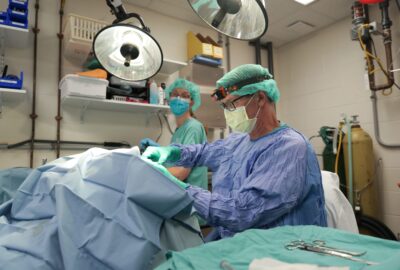
When most people think of X-rays, they picture a doctor’s or dentist’s office—wearing a heavy apron and waiting for the click of the machine. At the New England Aquarium, the same technology helps us care for a very different kind of patient. Our Animal Care team uses radiography to monitor the health of animals ranging from sea lions and penguins to sea turtles and anacondas.
Radiographs—the images created using X-rays—let us look beneath scales, feathers, and fur to diagnose injuries, infections, and other conditions that aren’t visible on the surface. They’re also an essential part of many animals’ annual exams, helping establish a baseline for what normal looks like for each individual. Those reference images make it easier to spot subtle differences over time, from early signs of arthritis to age-related bone changes, guiding care throughout an animal’s life.
How do we get our unique patients to cooperate for their close-up? That depends on the species.
Pinnipeds: Seals and sea lions
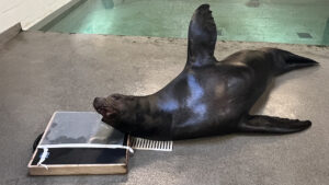
The Aquarium’s seals and sea lions are trained to participate voluntarily in radiographs. Through positive reinforcement and operant conditioning—providing rewards such as favored food items or enrichment activities—they learn to stay relaxed around each piece of equipment involved in the process. Trainers gradually introduce the animals to the X-ray stand, then the machine itself, the plate they rest on, and even the sight of staff wearing protective lead aprons.
Once trained, they’ll patiently scoot along the plate while the Animal Care team captures a full series of images, from head to hind flipper, including both top-down (dorsoventral) and side (lateral) views. Some sea lions even “open wide” for dental radiographs.
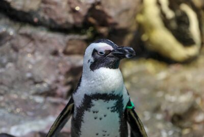
Penguins
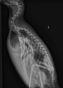
Penguins also participate in radiographs, though their setup looks a little different. We perform standing radiographs, which is exactly what it sounds like—the birds stand upright inside a custom acrylic box designed just for them. One side of the box slides open for easy entry and exit, and many penguins waddle right in when it’s their turn.
Once they’re comfortably in place, we capture two images: a front-to-back (ventrodorsal) view and a side view. Occasionally, we pause for a moment while a penguin shifts into position, but with a little patience, the results are almost picture-perfect.
Cold-stunned sea turtles
Each fall, hundreds of cold-stunned sea turtles arrive at the Aquarium’s Sea Turtle Hospital at our Animal Care Center in Quincy, MA, after washing ashore from frigid Cape Cod Bay.
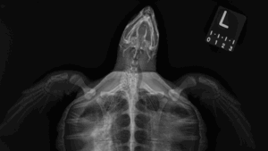
Every turtle receives intake radiographs upon arrival, which help Aquarium veterinarians diagnose injuries and illness. Cold-stunned turtles often develop pneumonia or, in some cases, bone infections such as osteomyelitis, both of which are visible on the radiographs.
Follow-up radiographs over the next several weeks allow veterinarians to track healing and monitor changes in the lungs and bones as the turtles recover. Amid the seriousness of treatment, the images sometimes reveal lighter details too, like bits of crab in a Kemp’s ridley sea turtle’s digestive tract, a favorite snack of theirs.
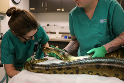
Anacondas
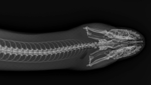
Capturing a clear radiograph of a snake takes patience and precision. Because even slight movement can blur the image, keeping them steady is essential.
To obtain good images of these naturally wiggly patients, we use light sedation to help them relax throughout the process. We may also use a snake tube, a clear device that allows the anaconda to rest safely inside while staying straight and still.
Much like with our seals and sea lions, the team takes a series of radiographs from head to tail, ensuring every inch of the lengthy patient is examined and recorded.
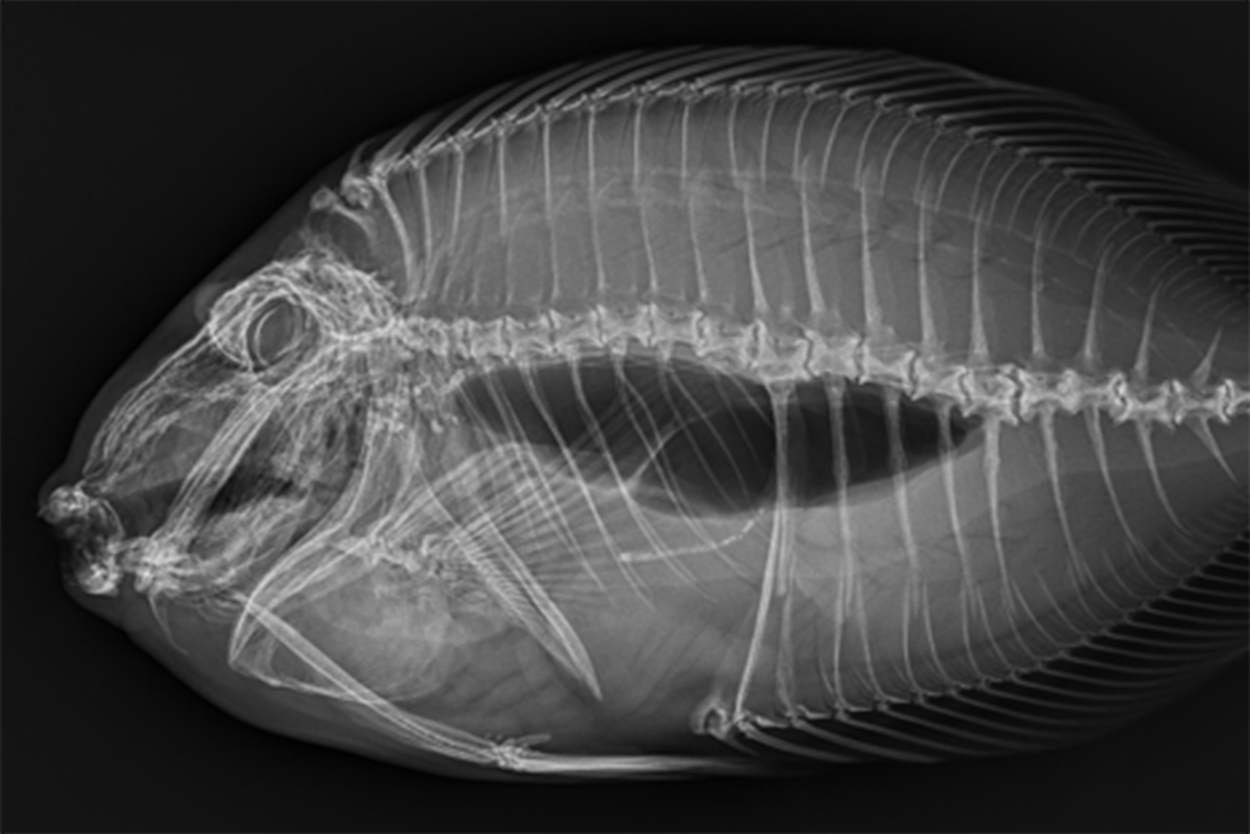
Fish
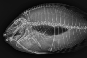
Fish sometimes receive radiographs too! To begin, they’re gently sedated in anesthetic water and moved quickly but safely to the X-ray table for imaging. Then, they are promptly returned to their anesthetic water for the rest of their exam.
We typically take a right lateral radiograph, meaning the fish’s right side rests flat against the radiology plate. On radiographs, dense structures like bones appear white while air-filled areas, such as the trachea or swim bladder, appear dark, revealing details about the fish’s anatomy and overall health.
We also take dorsoventral radiographs, positioning the fish in a shallow, water-filled trough so the patient remains supported during imaging from above. Capturing these orthogonal, or multiple-view, images allows veterinarians to see the body from different angles, making it easier to identify subtle changes or hidden issues.
These images may not hang in our galleries, but they’re some of the most important photographs we have—revealing knowledge that keeps animals at the Aquarium healthy and thriving.
Next time you visit the Aquarium, as you watch sea turtles swim and pinnipeds play, remember there’s a dedicated medical team behind the scenes caring for them every day!

Related Stories

Protect the Blue Planet
Your ticket purchase to the Aquarium directly supports our work to create a vital and vibrant ocean for future generations.